
Skin Lesion Removal in Brighton and Hove
Clinically assessed aesthetic removal of selected benign skin lesions, including skin tags, milia, cherry angiomas, seborrhoeic keratoses, warts, verrucae and selected benign moles.
Small skin lesions can be physically irritating, visually distracting or simply something you no longer want to keep noticing.
They may catch on clothing, bleed when shaving, interrupt the texture of your skin, or become the thing your eye goes to every time you look in the mirror.
At odNOVA Aesthetics, suitable benign lesions can be removed with a precise, clinically assessed approach designed to support neat healing and a smoother, more even skin appearance.
The odNOVA Standard: Careful removal starts with knowing what not to remove. Any lesion that appears suspicious, changing or uncertain is not treated cosmetically.

Dermoscopy-led assessment.
Each lesion is examined before treatment to assess whether cosmetic removal is appropriate.
Prices from £75
Smaller benign lesions start from £75. More complex lesions start from £100.
Local anaesthetic where needed.
Used where clinically appropriate and included in the treatment cost.
Minimal disruption.
Most treated areas heal with temporary redness, pinkness or small scabbing, depending on the lesion type and aftercare.
Aesthetic removal, clinical judgement
A small lesion does not need to be medically serious to be annoying. It may catch on clothing, bleed when shaving, interrupt the texture of your skin, or simply be the thing you keep noticing in the mirror.
At odNOVA Aesthetics, removal is approached with clinical discipline. The goal is not to “zap” everything in sight. The goal is to assess the lesion properly, choose the most appropriate method, and leave the smallest sensible footprint on the skin.
If a lesion appears suspicious, changing, atypical or diagnostically uncertain, I will not treat it as a cosmetic procedure. In that situation, I may recommend GP review, dermatology referral or histology.
Suitable benign lesions can often be removed quickly and neatly — but only after they have been assessed properly.
Your safety is not negotiable.

odNOVA Aesthetics mini PORTFOLIO
SEE RESULTS

Before treatment view of multiple benign skin tags on the neck area prior to aesthetic removal at odNOVA Aesthetics in Brighton. The lesions are clinically assessed via dermoscopy to ensure suitability for the procedure.

Multiple benign skin tags removed in a single session. The image shows the skin just 30 minutes after the procedure; early redness is a normal part of the initial healing phase and typically settles quickly.


Facial skin appearance 14 days after benign mole removal and a professional TCA peel. The results show a significantly smoother complexion with an even skin tone.

Before
After 5 days (healing in progress)
Skin Lesion Removal Prices
Treatment type
Smaller benign lesions
More complex lesions
Multiple lesions
Histology, if required
Price Guidance
from £75
from £100
Quoted after assessment
Discussed separately
Clinical policy:
Local anaesthetic is included where clinically appropriate. The consultation fee is applied in full towards treatment if the procedure is performed within 6 months. Final pricing depends on lesion type, number, size, location and treatment method.

Seborrhoeic keratoses can often grow quite large, developing a waxy, 'stuck-on' texture that can be very noticeable, especially near the hairline or temples.
In this case, I assessed the lesion using dermoscopy to ensure it was clinically consistent with a benign seborrhoeic keratosis. Because of its size and location, a precise removal technique was essential to minimise disruption to the surrounding skin.

This image shows early healing seven days after seborrhoeic keratosis removal. Mild pinkness can be normal at this stage, and final settling varies depending on lesion type, skin type and aftercare.

Patients often come to me with multiple concerns in one area. Using a precise, clinically-led approach, I addressed both the texture and the pigmentation.

The image shows visible improvement in texture and pigmentation during the healing phase. Final results vary depending on the lesion type, treatment method, skin type and aftercare.

Lesions on the neck and jawline can be a constant source of irritation, especially when they catch on clothing or interfere with daily routines. This case demonstrates the removal of a raised benign lesion that had become bothersome for the patient.

This is an immediate post-treatment appearance. A small protective crust can be part of normal early healing and should be allowed to settle naturally.
What to expect
1. Consultation and dermoscopy:
I examine each lesion to assess whether it appears benign and suitable for aesthetic removal.
2. Suitability decision:
I will discuss the most appropriate treatment method with you. If there is diagnostic uncertainty, I may recommend further clinical review.
3. Targeted removal:
Techniques are selected according to the lesion (e.g., electrocautery, shave removal, cryotherapy, or extraction).
4. Aftercare:
You will receive clear instructions to support healing and protect the treated area while the skin settles.

When to get checked
If you notice any of the following ABCDE warning signs, arrange prompt medical or dermatology assessment:
Asymmetry
one half looks different from the other
Border
edges are irregular, blurred, or notched
Colour
multiple shades or uneven pigmentation
Diameter
larger than 6 mm
Evolving
changes in size, colour, shape, or other symptoms (itching, bleeding)
Benign lesions I commonly assess and treat

Skin Tags / Acrochordons
What they are:
Skin tags are small, soft pieces of raised skin that often appear on the neck, underarms, eyelids, groin or areas of friction. They are usually harmless, but they can become physically irritating — catching on clothing or jewellery, bleeding when shaving, or simply becoming a visual distraction you no longer want to notice.
How I approach removal:
Before any procedure, I assess whether the lesion appears consistent with a benign skin tag and suitable for aesthetic removal. Some lesions can mimic skin tags, including viral warts or other skin changes, so assessment matters. Once suitability is established, I use a controlled method such as electrocautery, cryotherapy or another appropriate technique, aiming for the smallest sensible footprint on the skin.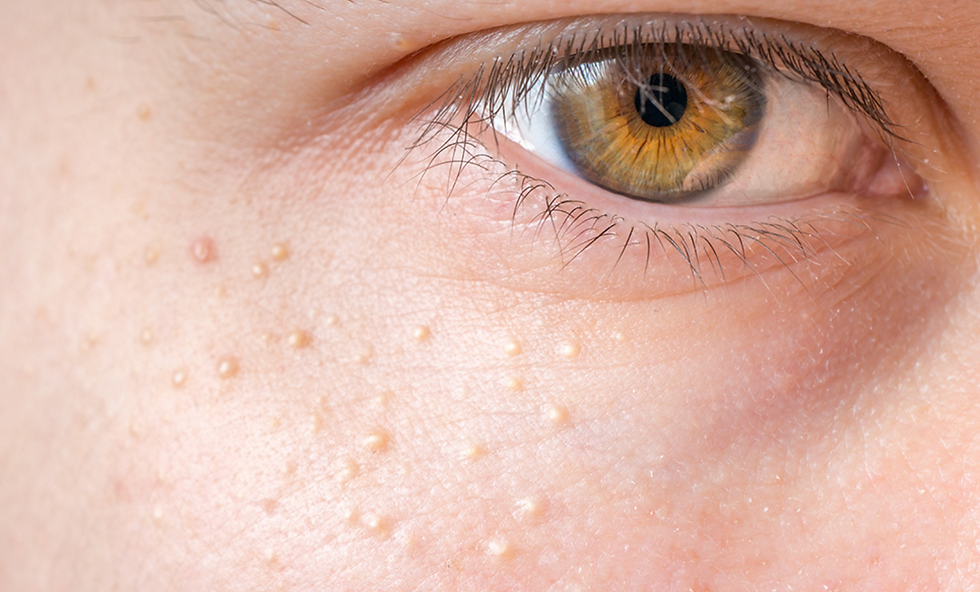
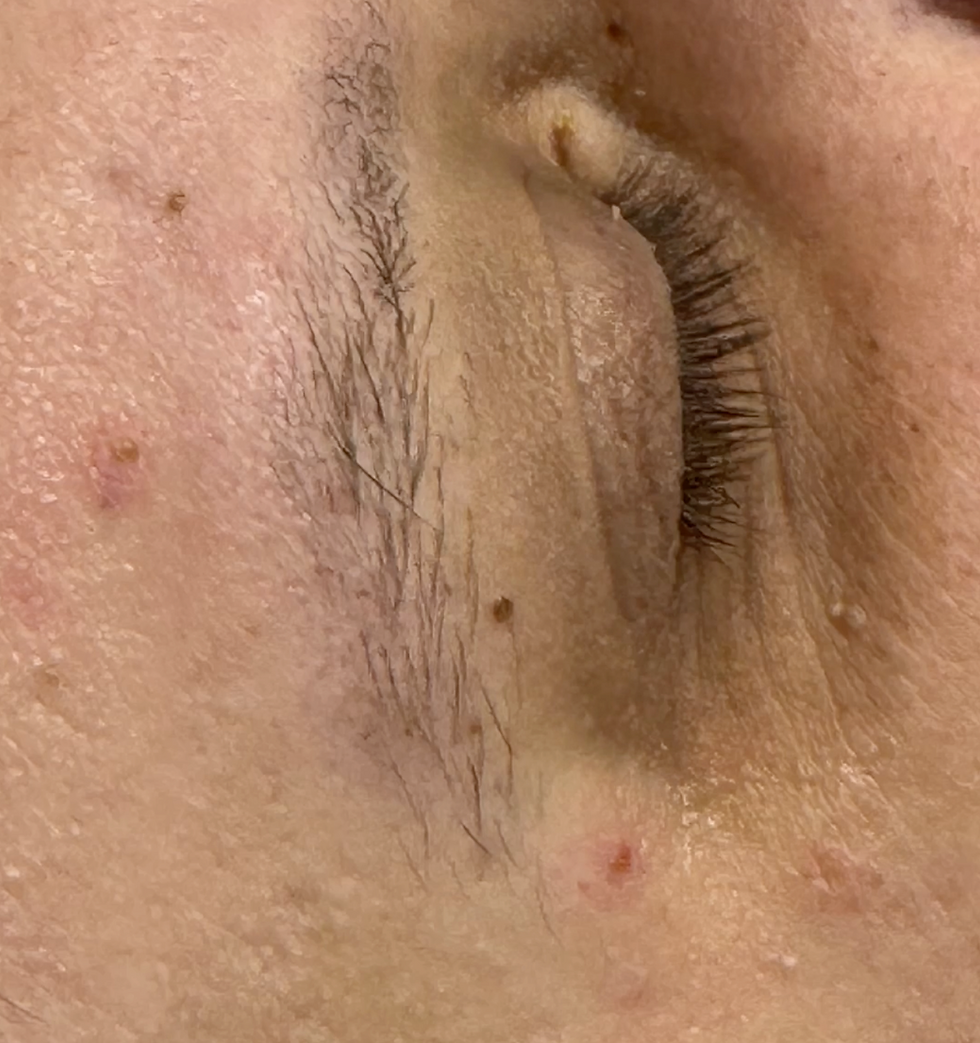
Milia
What they are:
Milia are tiny white or cream-coloured bumps that often appear around the eyes, cheeks or upper face. They sit under the surface of the skin and usually do not respond to squeezing, exfoliation or standard skincare. Trying to remove them at home can cause unnecessary trauma, redness or pigmentation.
How I approach removal:
I first assess whether the lesion appears consistent with milia and whether the location is safe to treat, especially around the eye area. Where suitable, milia can often be removed using careful professional extraction with minimal disruption to the surrounding skin. The aim is not aggressive squeezing — it is controlled release with a clean, precise technique.
Cherry Angiomas / Campbell de Morgan Spots
What they are:
Cherry angiomas are small red or ruby-coloured vascular spots that commonly appear on the trunk, arms, neck or face. They are usually harmless, but they can stand out visually, catch when shaving, or occasionally bleed if scratched or traumatised.
How I approach removal:
Before treatment, I assess whether the lesion appears consistent with a benign cherry angioma and suitable for aesthetic removal. Because these lesions are vascular, the treatment method needs to be controlled and precise. Where appropriate, I may use electrocautery or another suitable technique to treat the lesion while limiting unnecessary trauma to the surrounding skin.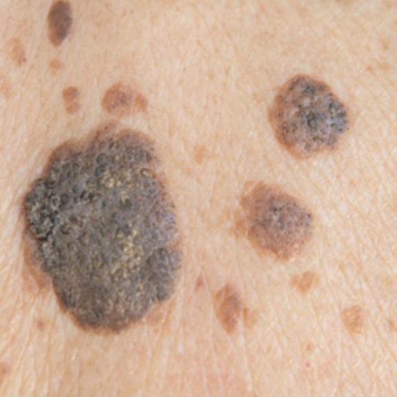
Seborrhoeic Keratoses
What they are:
Seborrhoeic keratoses are raised, rough, waxy or “stuck-on” looking lesions that often appear with age. They can be skin-coloured, brown, grey or darker, and may affect the texture of the skin. Some become itchy, irritated, crumbly or catch on clothing.
How I approach removal:
These lesions can sometimes resemble other pigmented skin changes, so assessment is essential before treatment. I assess the lesion clinically and, where appropriate, with dermoscopy to decide whether aesthetic removal is suitable. Depending on the thickness, texture, site and attachment to the skin, treatment may involve cryotherapy, curettage, electrocautery or another appropriate method.
Warts and Verrucae
What they are:
Warts and verrucae are viral lesions that can become rough, raised, thickened, persistent or uncomfortable. Verrucae commonly appear on the feet and may feel painful when walking. Warts can also affect the hands, face or other areas and may be cosmetically frustrating or difficult to clear.
How I approach removal:
I assess the lesion to check whether it behaves like a wart or verruca and whether treatment is appropriate. These lesions may require staged treatment rather than a single quick removal, especially if they are thick, longstanding or located on pressure areas. The method is selected according to the site, depth, thickness and behaviour of the lesion.
Solar Lentigines / Sun Spots
What they are:
Solar lentigines are flat brown marks that commonly appear on sun-exposed areas such as the face, hands, chest, shoulders and forearms. They are often associated with cumulative UV exposure and can make the skin look more uneven or aged.
How I approach removal:
Pigmented lesions require particular caution. Before any aesthetic treatment, I assess whether the lesion appears suitable and whether further review is needed. Some pigmented lesions can mimic more serious skin conditions, so cosmetic treatment is not appropriate if there is uncertainty. Where suitable, treatment is selected according to the pigment pattern, skin type, site and risk of post-inflammatory pigmentation.
Moles / Melanocytic Naevi
What they are:
Moles, also called melanocytic naevi, are common pigmented skin lesions made up of melanocytes — the pigment-producing cells of the skin. They may be flat or raised, skin-coloured, brown or darker, and can appear anywhere on the face or body. Some moles are present from childhood, while others develop over time.
How I approach removal:
Mole removal requires careful clinical assessment and dermoscopy before any aesthetic treatment is considered. I only consider removal when the mole appears clinically benign and suitable for aesthetic removal. If a mole is changing, irregular, symptomatic, unusually pigmented or diagnostically uncertain, cosmetic removal is not the correct first step. In that situation, I may recommend GP review, dermatology referral or histology.Where a mole appears suitable, the treatment method is chosen according to its size, depth, location and clinical features, with the aim of controlled removal and neat healing.
Expert Care
Skin lesion removal is performed by Piotr Wojtowicz, a pharmacist prescriber with advanced training in aesthetic medicine and postgraduate dermatology training.
I combine clinical science, dermoscopy-led assessment, ethics and precision to approach each lesion carefully before treatment.
No rushed “zap and go” approach. No treating what should not be treated cosmetically — just thoughtful, evidence-based care shaped around your skin, your concern and what is clinically appropriate.